In the field of the Dentistry, the digital workflow has evolved very fast. On this matter, when we are taking impression through digital protocols for complete Dentures, what is the best technique to take this kind of impressions?
In order to answer this question we have to consider the following:
1. Fundamental Limitation of intraoral scanners in complete dentures
Intraoral scanners (IOS) were developed primarily for tooth-supported, rigid reference structures. In edentulous patients, several critical challenges exist:
- Lack of stable landmarks for image stitching
- Mobile, compressible mucosa (especially in the vestibules and posterior palatal seal area)
- Difficulty capturing functional border movements
- Limited accuracy for peripheral extensions
As a result, direct digital impressions of edentulous arches using IOS alone remain inferior to conventional functional impressions in terms of denture retention and stability.
2. Current Gold Standard: Digitally Assisted Conventional Impression (Hybrid Workflow). This is the technique we suggested at WeCAD4You:
Step 1: Conventional Functional Impression (Analog)
The best and most predictable technique today remains:
- Custom tray fabrication (** but the custom Tray can be designed by Direct primary Digital impression using a good IOS).
- Border molding (green stick compound or heavy body PVS)
- Final impression using:
- Zinc oxide–eugenol (ZOE), or
- Light-body polyvinyl siloxane (PVS), or Polyether
This step ensures:
- Accurate recording of functional vestibular depth
- Proper posterior palatal seal
- Controlled tissue displacement
Step 2: Digitization of the Definitive Impression or Master Cast
Once the functional impression is obtained:
- The impression or poured cast is digitized using a laboratory scanner or a High performance IOS.
- This produces a high-accuracy STL file for CAD design
This approach combines:
- Biological accuracy (analog)
- Digital precision and reproducibility (CAD/CAM)
3. Alternative Digital Strategies (Emerging, but Not Yet Superior)
A. Direct IOS Impression of Edentulous Arches
Possible but case-selective:
- Best for well-keratinized, minimally mobile ridges
- Requires:
- Scanning in segments
- Artificial landmarks (scan sprays or markers)
- Still inferior for border accuracy
Clinical indication: Interim dentures, duplicates, or research settings.
B. Scan of Existing Well-Fitting Denture (Copy Denture Technique)
If the patient has a satisfactory old denture:
- Denture is scanned (intaglio + cameo surfaces)
- Used as a functional reference
- Can significantly improve digital outcomes
This is currently one of the most reliable digital shortcuts in complete denture therapy.
4. Recommended Best Technique (2025 Consensus)
Best clinical approach today:
Conventional functional impression using custom trays and border molding, followed by digital scanning and CAD/CAM denture fabrication.
This method:
- Respects prosthodontic biology
- Maximizes retention and stability
- Integrates seamlessly into modern digital workflows
- Has the strongest clinical validation
Practical Takeaway for Digital Workflow in Complete Dentures:
Digital dentistry enhances efficiency and reproducibility, but in complete dentures, biomechanics and soft-tissue management remain decisive. Until IOS technology can reliably capture functional soft tissues, hybrid workflows remain the standard of care.
Figures:
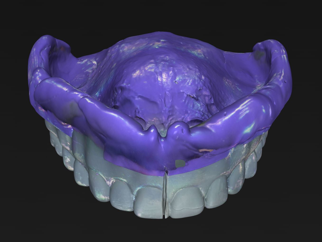
- An overextended primary impression obtained using a digital protocol does not accurately capture the peripheral seal area required for the design and manufacture of a complete removable prosthesis.
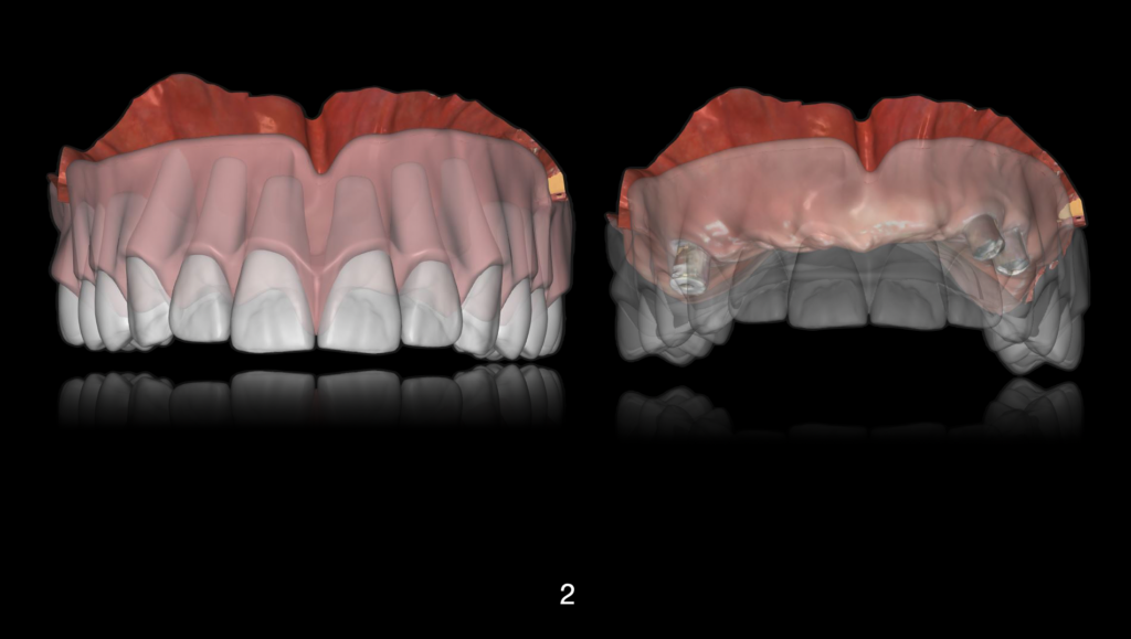
2. Under optimal circumstances, the primary impression may be used for the fabrication of a trial prosthesis (try-in). Based on this, primary esthetic and functional parameters can be evaluated.
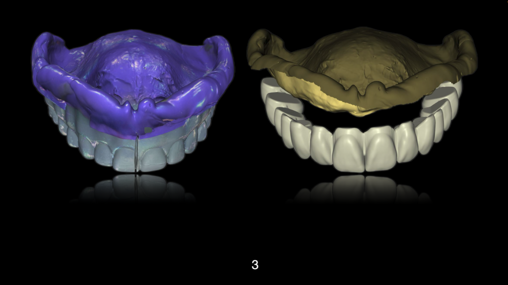
3. Using the trial prosthesis or try-in, it is possible to obtain a functional impression by means of conventional protocols involving mechanical border trimming and functional peripheral muscle insertion recording, as well as posterior palatal seal registration, through The functional impression.
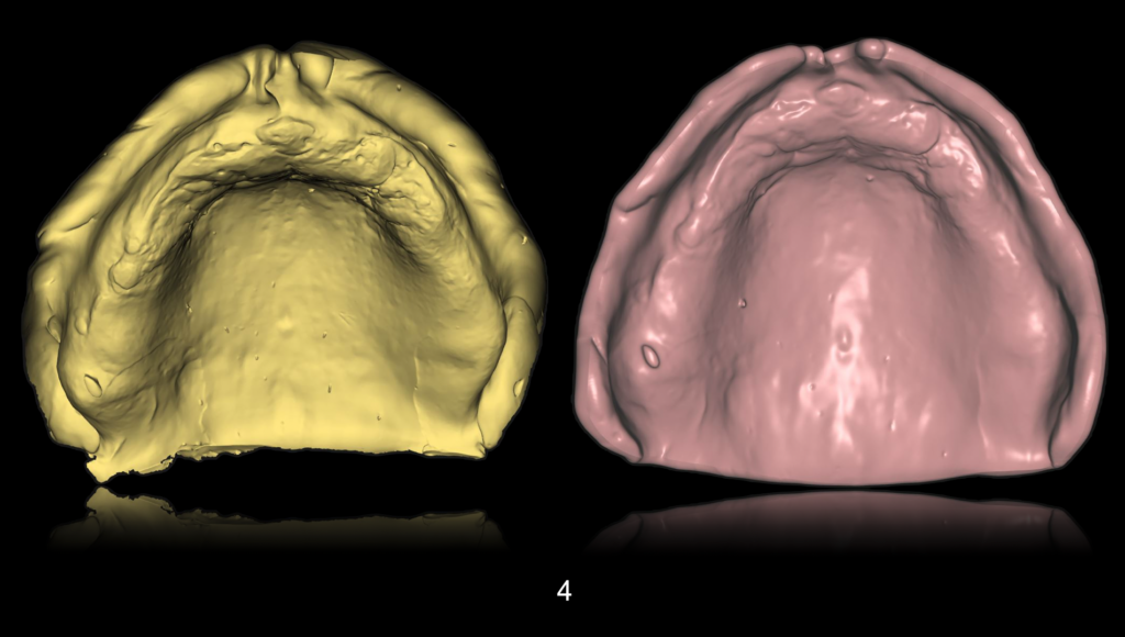
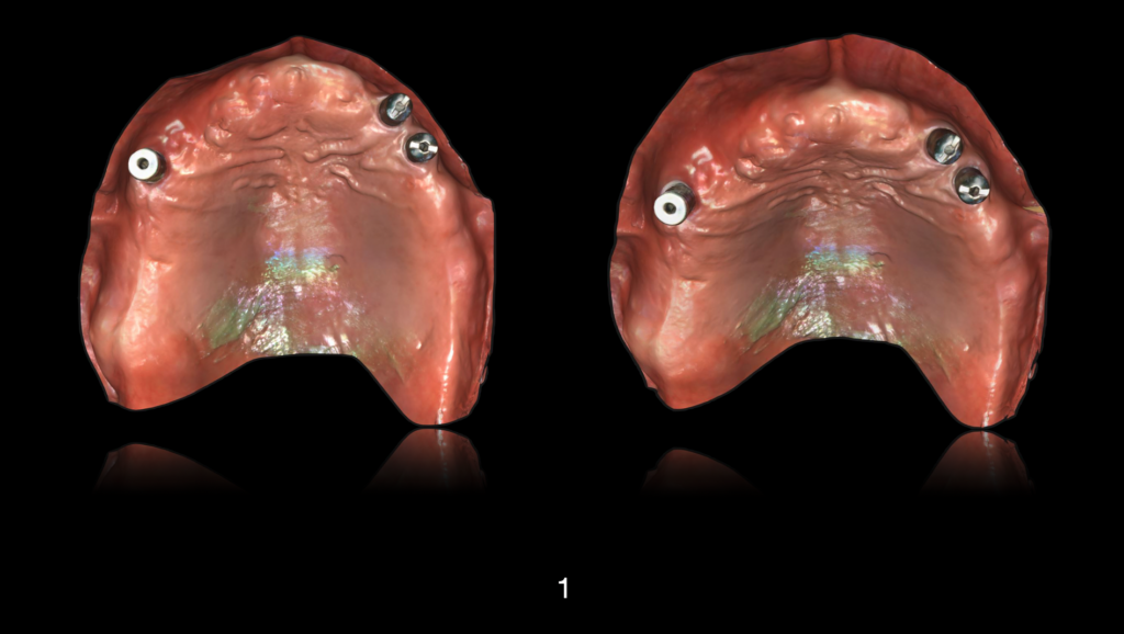
4. Within the digital workflow, through 360° digitization using high-end intraoral or laboratory scanners, it is possible to capture the peripheral seal area and transfer it to the design of the prosthetic base.
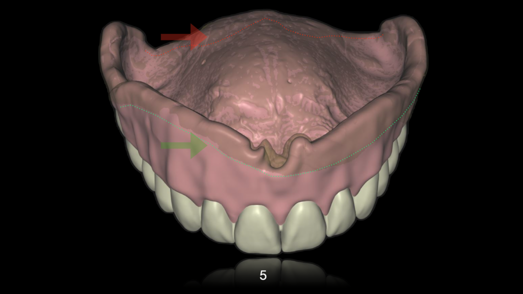
5. In this image, the critical areas captured in the analog impression and subsequently digitized through a mixed digital workflow protocol are delineated.
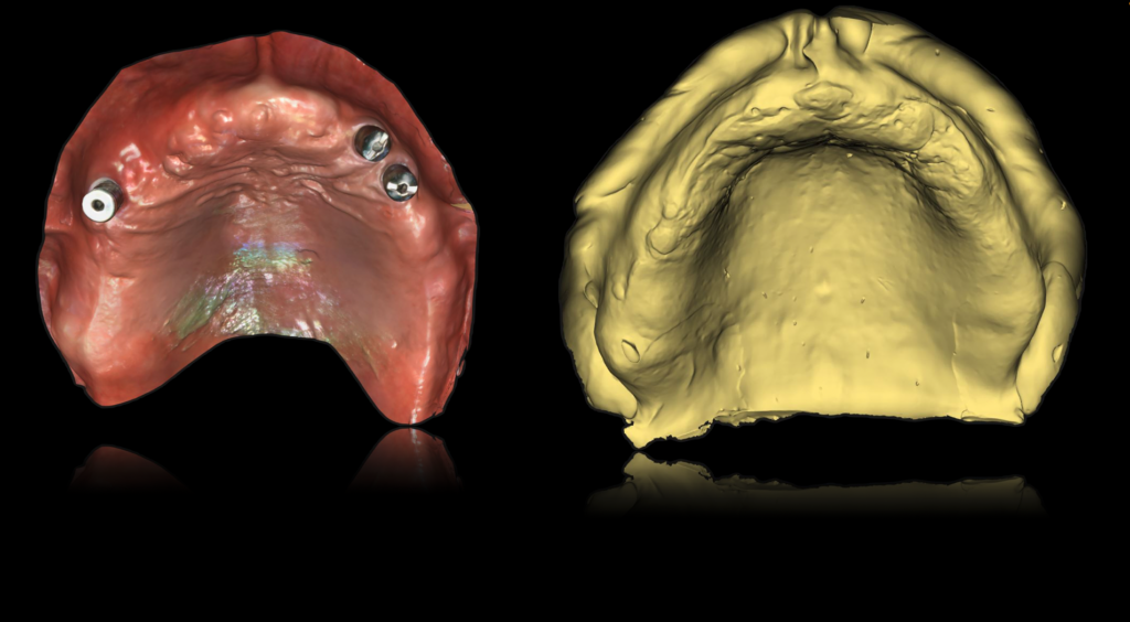
6. The success of the prosthetic appliance is based on accurate diagnosis and therapeutic planning, together with the selection of the most appropriate and reliable primary and functional impression protocols.
5. Key References
1. Goodacre BJ, et al.
Clinical complications in complete dentures.
J Prosthet Dent. 2011.
2. Srinivasan M, et al.
CAD/CAM complete dentures: a systematic review.
Clin Oral Investig. 2021.